CASE 36
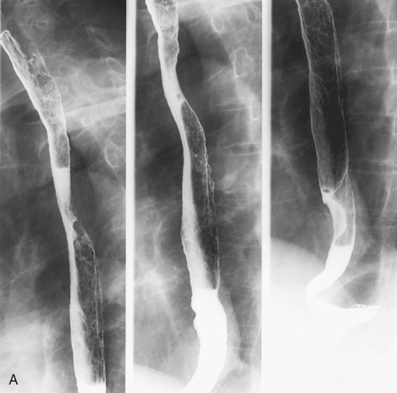
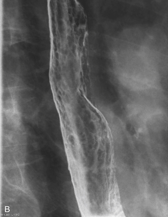
History: An 80-year-old man with oxygen- and steroid-dependent chronic obstructive airway disease develops dysphagia and odynophagia.
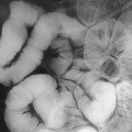
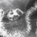
1. Which of the following should be included in the differential diagnosis of the dominant imaging finding on figure A? (Choose all that apply.)
D. Cytomegalovirus (CMV) esophagitis
2. Which of the following distinguishes esophageal Candida infection in an immunocompromised patient from the same occurring in an immunocompetent host?
D. The infection is identical in both the immunocompromised and the immunocompetent patient.
3. Which of the following statements about esophageal Candida infection is true?
A. Candida albicans is the most common cause of infectious esophagitis.
B. Most of the patients with Candida esophagitis also have clinically visible oropharyngeal thrush.
Stay updated, free articles. Join our Telegram channel

Full access? Get Clinical Tree


