The Deep Venous System I: Internal Cerebral Veins, Tributaries, and Drainage
36.1 Case Description
36.1.1 Clinical Presentation
A 64-year-old woman presented with seizures and progressive headaches. Outside computed tomography revealed a large mass centered at the tentorial incisura, and she presented to neurosurgery.
36.1.2 Radiologic Studies
See ▶ Fig. 36.1, ▶ Fig. 36.2, and ▶ Fig. 36.3.
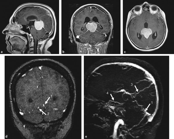
Fig. 36.1 In this patient with a tentorial incisura meningioma (a–c; T1-weighted postcontrast MRI), the major question to be answered before surgical removal is whether the straight sinus is functional and, if not, how the deep brain drains, as this will determine which veins to spare during surgery and which approach to take. Coronal MR venography (d) with sagittal reconstructions (e) suggests occlusion of the vein of Galen with a straight sinus stump (1), persistence of a left-sided tentorial sinus (2), superior drainage into a medial parietal vein (3), and anterior drainage of the right basal vein of Rosenthal (4). Digital subtraction angiography was performed to further elucidate these findings. Case continued in ▶ Fig. 36.2.

Fig. 36.2 Right internal carotid artery injection lateral (a) and AP (b) in the venous phase demonstrates drainage of the ICVs, rather than in the vein of Galen, craniomedially into two medial parietal veins (2) toward the superior sagittal sinus. The basal vein of Rosenthal is seen to drain laterally via the inferior temporal vein (4) toward the transverse sinus. The small white arrows denote the collateral pathways. Case continued in ▶ Fig. 36.3.
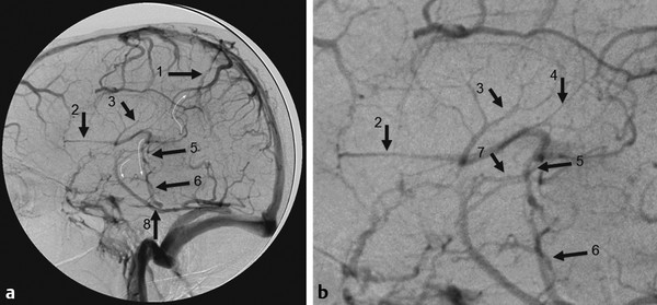
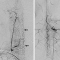
Fig. 36.3 Left internal carotid artery injection in the venous phase in lateral view (a, enlarged view in b) demonstrates drainage to the medial parietal veins (1) toward the superior sagittal sinus, but also drainage from the septal veins (2), the thalamostriate veins (3), and the posterior atrial veins (4) into the ICVs, and from there, via an inferior thalamic vein (5) to the lateral mesencephalic vein (6), which opens to the superior petrosal vein. This “epsilon” shape of the venous drainage is typically seen in vein of Galen AVMs, where the deep venous system has not gained access to a normally developed straight sinus drainage. Additional drainage of the basal vein of Rosenthal (7) in the present case is through the inferior temporal vein (8). The small curved white arrows denote the collateral pathways.
36.1.3 Diagnosis
Tentorial incisura meningioma with occlusion of the straight sinus and rerouting of the deep venous drainage.
36.2 Embryology and Anatomy
Related posts:


Stay updated, free articles. Join our Telegram channel

Full access? Get Clinical Tree


