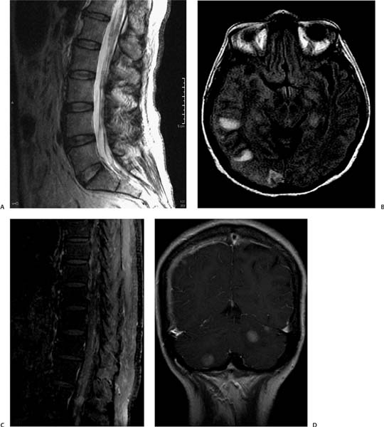
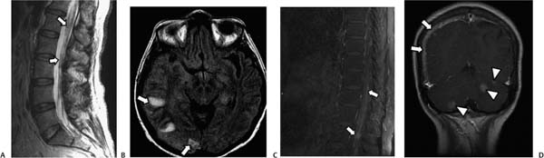
Case 37 A 60-year-old woman with the history of a lump in her left breast. (A) Sagittal T2-weighted image (WI) of the lumbar spine shows nodular thickening of the nerve roots (arrows). (B) Axial fluid-attenuated inversion recovery (FLAIR) image of the brain shows three foci of increased signal in the subcortical white matter on the right (arrows). (C) Sagittal T1WI of the spine after contrast with fat saturation shows diffuse nodular enhancement of the nerve roots (arrows). (D) Coronal T1WI of the brain with contrast shows thick meningeal enhancement on the right (arrows) along with foci of nodular enhancement in the posterior fossa (arrowheads). • Meningeal carcinomatosis:
Clinical Presentation
Imaging Findings
Differential Diagnosis
![]()
Stay updated, free articles. Join our Telegram channel

Full access? Get Clinical Tree


