 Clinical Presentation
History withheld.

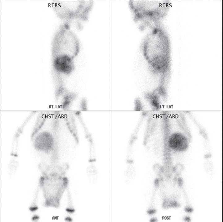
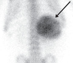
Tc99m HDP bone scintigraphy demonstrates intense growth plate activity and a large head-to-body size ratio, indicating that this is a young child. A large, markedly increased focus is seen in the right upper abdominal soft tissues. It is more intense on the posterior view, indicating that it is more posteriorly located (arrow). No abnormal bone foci are seen.
 Differential Diagnosis
• Primary neuroblastoma (NB): Soft-tissue uptake on bone scan in a posterior abdominal mass in a young child makes this the most likely diagnosis.
• Liver metastasis: Some liver metastases can be seen focally
Stay updated, free articles. Join our Telegram channel

Full access? Get Clinical Tree


