 Clinical Presentation
A 10-month-old infant with persistent cough and stridor.
Further Work-up
 Imaging Findings
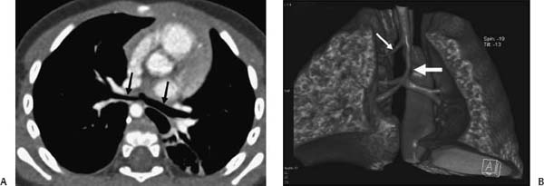
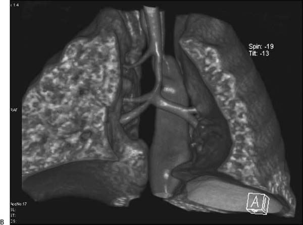
(A, B) Computed tomography (CT) of the chest: axial image at the level of the carina (A) and three-dimensional volume-rendered image (B) show an abnormally long segment of narrowing in the distal tracheal (thick white arrow), a horizontal orientation of the bronchi arising from the carina (black arrows), and a tracheal bronchus for the right upper lobe (thin white arrow).
 Differential Diagnosis
• Long-segment congenital tracheal stenosis:
Stay updated, free articles. Join our Telegram channel

Full access? Get Clinical Tree


