CASE 37
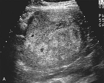
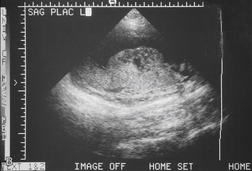
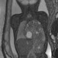
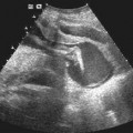
History: Two images of placentas are presented. Figure A is from one asymptomatic pregnant patient, and Figure B is from a second asymptomatic pregnant patient.
1. What should be included in the differential diagnosis for the placental lesion shown in Figure A? (Choose all that apply.)
B. Hematoma
2. Which lesion occurs more commonly on the maternal side?
B. Hematoma
3. Which laboratory abnormality is associated with the above-mentioned placental abnormalities?
A. Anemia
C. Leukocytosis
4. What is the diagnosis of the placental mass shown in Figure B?
Stay updated, free articles. Join our Telegram channel

Full access? Get Clinical Tree


