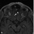
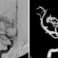
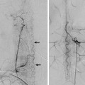
Fig. 37.1 T2-weighted axial images of a brain MRI (a,b) and conventional angiography (right internal carotid artery injection; anteroposterior [AP] view, early and late phase [c,d]) show a right temporal AVM nidus draining mainly via the deep middle cerebral vein (small arrows) into the right basal vein of Rosenthal (BVR). The posterior communicating vein (arrow in d) and the contralateral BVR are also enlarged and open into the left superior petrosal sinus, via the left lateral mesencephalic vein. This drainage pattern explains the left pulsatile tinnitus, even though the AVM is located in the right hemisphere.
37.1.3 Diagnosis
Right temporal arteriovenous malformation (AVM) draining through the venous circle of Trolard.
37.2 Anatomy
The basal vein of Rosenthal (BVR) originates at the junction between the anterior cerebral vein and the deep middle cerebral vein, just anterior to the midbrain near the anterior perforated substance. It courses superior and medial to the posterior cerebral artery around the midbrain, traversing the prepeduncular, ambient and quadrigeminal cisterns to finally open into the vein of Galen. In some cases, it may drain into the straight sinus or the internal cerebral veins. Alternatively, the BVR may be discontinuous, with the anterior part draining anteriorly to the cavernous sinus or posteriorly to the superior petrosal sinus via the lateral mesencephalic vein, and the posterior part to the vein of Galen. In addition, the BVR may keep its “embryonic” pattern and drain to a tentorial sinus, or even a temporal cortical vein.
The BVR is a venous efferent that forms late during embryogenesis and serves as a longitudinal anastomosis between telencephalic, diencephalic, and mesencephalic veins, serving as a connection between the deep middle cerebral vein and the vein of Galen. As the BVR is primarily not involved in the drainage of the subependymal venous network, embryologically speaking it cannot be regarded as part of the deep venous system, even though it is located in the depth of the brain. It anastomoses medially with its contralateral homolog through the venous circle (see following), and inferiorly with the petrosal vein via the lateral mesencephalic vein. Each of these channels constitutes an alternative outlet in case of venous obstruction or venous overflow (as seen in venous thrombosis or in regional AV shunts). The relatively late appearance of the BVR and the numerous tributaries account for the multiple variations encountered in the basal vein. The extent of the venous territories connecting to the BVR accounts for its unfavorable hemodynamic effects when it participates in the drainage of an AV shunt. Its drainage territory encompasses the orbital surface of the frontal lobe, the insula and mesial temporal lobe structures, the hypothalamus, parts of the striatum and thalamus, and the midbrain.
The venous circle, or circle of Trolard, connects the anterior afferents to the BVR in the chiasmatic region anteriorly and the ventral midbrain region posteriorly. Both anterior cerebral veins are connected medially by the anterior communicating vein, present in ~50% of the population (▶ Fig. 37.2). This vein parallels the anterior communicating artery as it courses along the lamina terminalis above the optic chiasm. It may have a plexiform configuration, with multiple small channels rather than a single vessel. The medial afferent branches that contribute to the anterior venous circle include the septal veins, callosal veins, anterior cerebral veins, chiasmatic veins, and olfactory veins near the midline. The lateral afferents include the inferior striate veins, insular veins, and uncal veins.
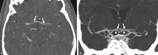
Fig. 37.2 The anterior communicating vein (arrows) in a patient being investigated for stroke with CTA (axial [a] and coronal [b] reformats) is seen to run in close spatial relation to the A1 segments (arrowheads) along the lamina terminalis and above the optic chiasm.
Related posts:


Stay updated, free articles. Join our Telegram channel

Full access? Get Clinical Tree


