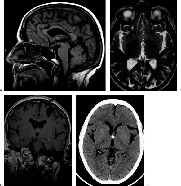
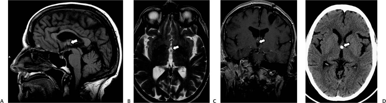
Case 38 An adult woman with severe intermittent headaches. (A) Sagittal T1-weighted image (WI) shows a round mass in the roof of the 3rd ventricle (arrow). The mass is isointense to the gray matter. (B) Axial T2WI shows the lesion as isointense to cerebrospinal fluid (arrow). (C) Coronal T1WI with contrast shows the lesion in the roof of the 3rd ventricle (arrow); there is no enhancement. (D)
Clinical Presentation
Imaging Findings
![]()
Stay updated, free articles. Join our Telegram channel

Full access? Get Clinical Tree


