 Clinical Presentation
A 53-year-old man with hemoptysis.
 Imaging Findings
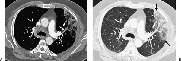
(A, B) Contrast-enhanced computed tomography (CT): axial images in mediastinal (A) and lung (B) windows demonstrate a pleura-based triangular opacity extending from the right hilum (black arrows), as well as the absence of contrast opacification in a segmental branch of the left upper lobe pulmonary artery (white arrow).
 Differential Diagnosis
• Pulmonary infarct secondary to acute pulmonary embolism (PE):
Stay updated, free articles. Join our Telegram channel

Full access? Get Clinical Tree


