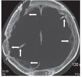
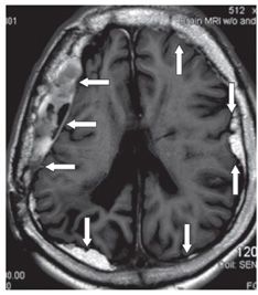
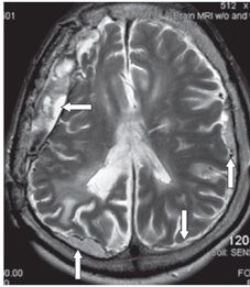
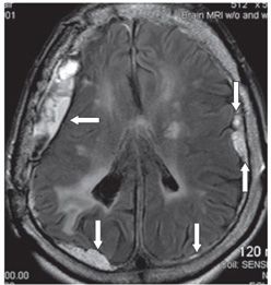
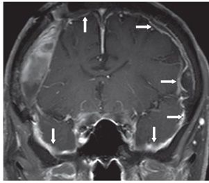
FINDINGS Figure 38-1. Axial NCCT through the centrum semiovale. There is a mixed density right frontal extraaxial collection (right transverse arrow). There is an area of focal hyperdensity (calcification/ossification) at the anterior end of the collection (vertical arrow). Left-sided dural bony excrescences are present (transverse left arrow). Figure 38-2. Axial NCCT bone window setting through the lateral ventricles. There is a left anterior transfrontal and a right posterior transparietal ventriculoperitoneal (VP) shunts (vertical arrows). Ossified excrescences are present along the inner table bilaterally with ossification of the falx (transverse arrows). Figures 38-3 to 38-5
Stay updated, free articles. Join our Telegram channel

Full access? Get Clinical Tree


