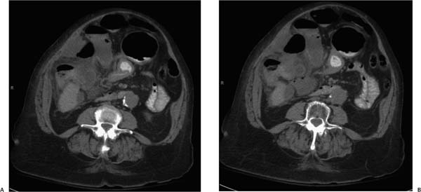
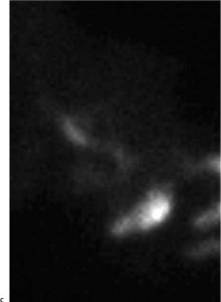
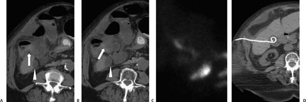
Case 38 An 82-year-old presents with right upper quadrant pain. (A,B) Axial computed tomography images show infiltration of the fat surrounding the hepatic flexure (arrows), liver, and gallbladder. The fundus of the gallbladder, seen posteriorly (arrowheads), appears normal, but the body shows marked mural thickening. (C) Hepatobiliary iminodiacetic acid (HIDA) scan demonstrates nonfilling of the gallbladder, consistent with acute cholecystitis. (D) Successful treatment by cholecystostomy tube placement.
 Clinical Presentation
Further Work-up
 Imaging Findings
 Differential Diagnosis
Stay updated, free articles. Join our Telegram channel

Full access? Get Clinical Tree


