The Infratentorial Veins
38.1 Case Description
38.1.1 Clinical Presentation
A 39-year-old man presented with a long-standing history of headaches. CTA revealed a right cerebellar arteriovenous malformation (AVM) nidus. He was referred for elective angiogram.
38.1.2 Radiologic Studies
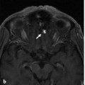
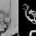
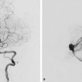
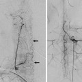
See ▶ Fig. 38.1.
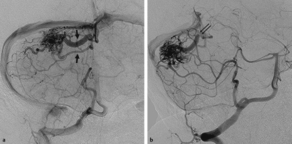
Fig. 38.1 Right vertebral artery angiograms in anteroposterior (AP) (a) and oblique (b) views demonstrate a compact right cerebellar AVM nidus with supply from both the posterior inferior cerebellar artery and the superior cerebellar artery. There are two superior hemispheric veins draining toward the torcular (arrows). The more dilated draining vein shows a focal narrowing before entering the torcular (thin double arrows).
38.1.3 Diagnosis
Cerebellar AVM with drainage toward the torcular and focal stenosis of one of the draining veins.
38.2 Anatomy
The infratentorial veins can be divided into four separate groups: the superficial, deep, brainstem, and bridging veins.
38.2.1 Superficial Veins
The superficial infratentorial veins drain the cortical surfaces of the cerebellum and are grouped according to the cerebellar surface they drain (tentorial, petrosal, or suboccipital) and whether they are involved in the venous drainage of the hemisphere or the vermis.
The tentorial surface drains via two main veins: the superior vermian and the superior hemispheric veins. The superior vermian veins drain the superior vermis and medial aspect of the tentorial surface of the cerebellar hemispheres. They are subdivided in two groups: the anterior group, which will ultimately drain into the vein of Galen via the superior vermian vein proper and that courses through the quadrigeminal cistern, and the posterior group, which will ultimately drain into the torcular. The latter can drain into the torcular alone or after joining the inferior vermian vein. The superior hemispheric veins are further subdivided into an anterior and a posterior group and a much smaller lateral group. The anterior group will drain via the cerebellomesencephalic fissure vein or the superior vermian vein. The posterior group drains into the torcular, superior petrosal, transverse, or tentorial sinuses via bridging veins. They usually form a common trunk with the inferior hemispheric veins from the suboccipital surface before draining into the sinuses. The lateral group drains directly or indirectly into the superior petrosal sinus.
The suboccipital surface drains via three main veins: the inferior vermian, the inferior hemispheric, and the superficial tonsillar veins. The inferior vermian vein is usually a paired structure that runs along the cerebellovermian fissure and drains to the torcular or the transverse sinus directly or indirectly via a tentorial sinus. The inferior vermian veins drain the inferior vermis and medial aspect of the suboccipital surface of the cerebellar hemispheres. The inferior hemispheric veins can run longitudinally or transversely. Most of the longitudinal veins ascend to join the superior hemispheric veins and subsequently empty into a dural or tentorial sinus. The transverse veins are in relation to the cerebellar fissures, and most of them drain medially into the inferior vermian vein. The superficial tonsillar veins are subdivided into retrotonsillar, medial, and lateral groups. The medial and lateral veins drain into the retrotonsillar veins, which subsequently converge to form the inferior vermian veins.
The petrosal surface is drained mainly by anterior hemispheric veins. These are subdivided into superior, middle, and inferior groups, which converge in the region of the flocculus to form the vein of the cerebellopontine fissure. This vein will ultimately drain into the superior petrosal sinus.
38.2.2 Deep Veins
The main deep veins run longitudinally and are located within the three fissures in the cerebellum. They are divided into two groups: the veins of the fissures and the veins of the cerebellar peduncles.
The veins running along the fissures include the cerebellomesencephalic vein, cerebellopontine vein, and cerebellomedullary vein. They act mainly as collectors for the cerebellar hemispheric veins and the veins of the cerebellar peduncles.
The veins of the superior and inferior cerebellar peduncles run along the posterior surface of their respective peduncles, within the cerebellomesencephalic and cerebellomedullary fissures. The vein of the middle cerebellar peduncle runs on the lateral surface of the peduncle in the anterior part of the cerebellopontine fissure.
38.2.3 Veins of the Brainstem
For conceptual purposes, the veins of the brainstem can be divided according to the segment they drain and the direction in which they course.
Stay updated, free articles. Join our Telegram channel

Full access? Get Clinical Tree


