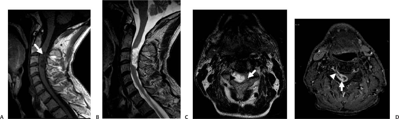
Case 39 A 27-year-old woman with progressive paresthesia in the right shoulder and arm. (A) Sagittal T1-weighted image (WI) of the cervical spine shows a low-signal lesion at the level of C3 (arrow). The lesion is extra-axial. (B) Sagittal T2WI of the cervical spine shows high signal of the mass. (C) Axial T2WI shows the mass (arrowhead) in the right aspect of the intradural space. The lesion compresses the spinal cord (arrow). (D) Axial fat-saturated T1WI of the cervical spine with contrast shows the “target” pattern of enhancement of the intradural, extramedullary mass (arrow). The lesion extends to the right neural foramen (arrowhead). • Spinal schwannoma:
Clinical Presentation
Imaging Findings
Differential Diagnosis
![]()
Stay updated, free articles. Join our Telegram channel

Full access? Get Clinical Tree


