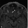
Fig. 39.1 T2-weighted (a,b,e) and contrast-enhanced T1-weighted (c,d) sagittal (a,b,c) and axial (d,e) MRIs demonstrate an intradural intramedullary arteriovenous malformation (AVM) in addition to a paravertebral AVM with multiple dilated vessels within the left lateral pedicle of T12 and the paraspinal tissues, including the musculature at this level on the left. Magnetic resonance angiography (f) demonstrates extensive shunting in both the cord and the paraspinal soft tissues centered on the T12 level. Case continued in ▶ Fig. 39.2.
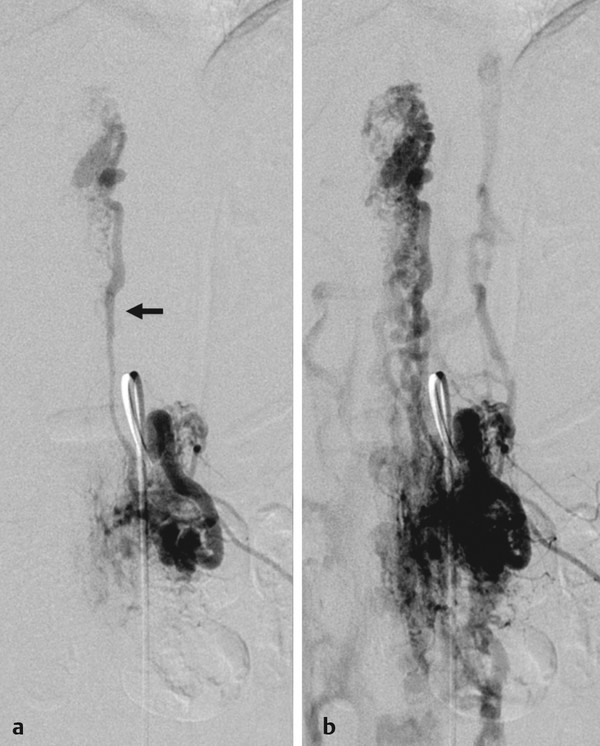
Fig. 39.2 Spinal angiography in the anteroposterior view in early (a) and late (b) arterial phases after injection into the left T12 segmental artery reveals the paravertebral AVM with arteriovenous shunting along the segmental artery supplying the skin, paravertebral muscles, and vertebral body, as well as the origin of a dorsolateral radiculopial artery (arrow) that supplies a spinal cord AVM. The concurrence of arteriovenous malformations affecting multiple compartments of the same metamere gave rise to the term spinal arteriovenous metameric syndrome.
39.1.3 Diagnosis
Spinal arteriovenous metameric syndrome (Cobb syndrome or juvenile spinal arteriovenous malformation) at T12.
39.2 Anatomy
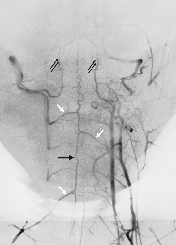
The blood supply to a given metamere (which consists of the vertebral body, the paraspinal muscles, the dura, nerve root, and spinal cord) is derived from segmental arteries. These segmental arteries are present in the fetus for each of the 31 spinal segments. This segmental supply remains preserved in the thoracic and lumbar regions via the intercostal or lumbar arteries. In the upper thoracic region, several segmental arteries coalesce to form a common feeder, which is the supreme intercostal artery. In the cervical region, this vascular rearrangement is even more obvious: on each side, three longitudinal anastomotic arteries are established as potential sources of spinal blood supply; namely, the vertebral artery, the deep cervical artery, and the ascending cervical artery.
The vertebral artery is a chain of intersegmental anastomoses that connects the cervical segmental arteries, each of which is able to supply a cervical segment, with the most prominent being the arcade of the dens. In the upper cervical region, potential sources of metameric blood supply are via anastomoses to the external carotid artery, mainly the occipital artery (namely, the C1 and C2 anastomoses; see Case 4) and the ascending pharyngeal artery (namely, the hypoglossal artery, which anastomoses with the C3 collateral of the vertebral artery via the odontoid arterial arch that supplies the dens; see Case 30). In the sacral and lower lumbar region, sacral arteries and the iliolumbar artery (which often supply the L5 level) derived from the internal iliac arteries are the most important supply to the caudal spine.
In general, the segmental arteries supply all the tissues on one side of a given metamere, with the exception of the spinal cord. Because of its embryological origin, each metamere is centered at the level of the vertebral disk, and therefore, each vertebra is supplied by two consecutive segmental arteries on both sides that anastomose extensively both across the midline and between levels above and below. The latter is formed by an extraspinal longitudinal system that connects the neighboring segmental arteries longitudinally. The vessels course on the lateral aspect of the vertebra or transverse process. This system is highly developed in the cervical region, where the vertebral artery and the deep cervical and ascending cervical arteries form the most effective chain of longitudinal anastomoses (▶ Fig. 39.3).
