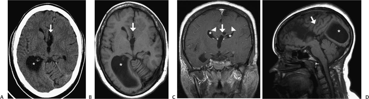
Case 4 A 12-year-old boy with slight cognitive defects. (A) Axial computed tomography (CT) scan of the head shows an absence of the anterior fibers of the corpus callosum (arrow) and a dilated lateral ventricle on the right (asterisk). (B) Axial T1-weighted magnetic resonance (MR) image of the brain shows an absence of the anterior fibers of the corpus callosum (arrow) and a dilated lateral ventricle on the right (asterisk). (C) Coronal T1-weighted image (WI) of the brain after intravenous contrast shows absence of the corpus callosum with a high-riding 3rd ventricle (arrow). Note the “bullhorns” appearance of the lateral horns (asterisks). Thick bands of white matter superior to the lateral ventricles are Probst fibers (arrowheads). (D) Sagittal T1WI of the brain shows absence of the corpus callosum. Note the lack of a cingulate gyrus, the vertically oriented sulci (arrow), and the colpocephaly of the right lateral ventricle (asterisk). • Dysgenesis of the corpus callosum (CC):
Clinical Presentation
Further Work-up
Imaging Findings
Differential Diagnosis
![]()
Stay updated, free articles. Join our Telegram channel

Full access? Get Clinical Tree


