 Clinical Presentation
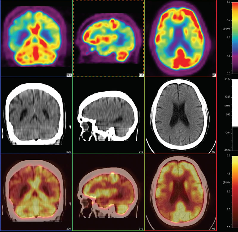
A 63-year-old woman with memory loss.

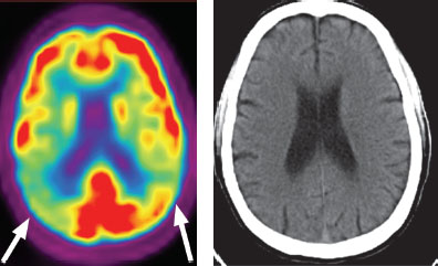
Selected axial, sagittal, and coronal images from an FDG-PET/CT of the brain demonstrate nearly symmetric, markedly decreased glucose metabolism of the posterior temporal and parietal lobes (arrows). CT shows mildly prominent ventricles for age, but findings are otherwise normal.
 Differential Diagnosis
• Alzheimer dementia (AD): Nearly symmetric involvement of the posterior temporoparietal cortices with sparing of the occipital lobes, basal ganglia, and cerebellum is the typical appearance of this diagnosis, as shown here.
• Lewy body dementia (DLB): This is the second most common degenerative dementia after AD. It can look similar to AD but typically has more pronounced occipital involvement.
• Multi-infarct dementia (MID):


Stay updated, free articles. Join our Telegram channel

Full access? Get Clinical Tree


