 Clinical Presentation
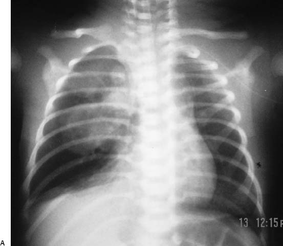
A 3-week-old infant with respiratory distress and decreased breath sounds in the right hemithorax.
Further Work-up
 Imaging Findings
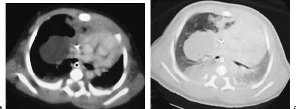
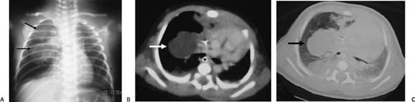
(A) Conventional radiograph shows a well-defined lobulated mass in the right middle and upper lung zones (arrows). (B, C) Computed tomography with mediastinal and lung windows confirms the presence of the low-density mass (arrows) and atelectasis of the right lower and middle lobes.
 Differential Diagnosis
• Congenital cystic adenomatoid malformation (CCAM) of the lung: In a newborn or a young infant, CCAM of the lung may present as a cyst or mass with a variable degree of compression and mass effect on the adjacent lung and mediastinum.
• Bronchogenic cyst: Bronchogenic cyst may also manifest early in infancy, and the presentation may include either a mediastinal or a pulmonary mass or cyst.
• Lymphangioma/cystic hygroma:
Stay updated, free articles. Join our Telegram channel

Full access? Get Clinical Tree


