Courtesy of Terri Love MD.

Courtesy of Terri Love MD.

Courtesy of Terri Love MD.
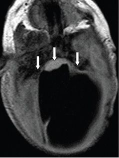
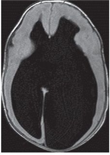
FINDINGS Figure 4-1. Axial T2WI through foramen magnum. Large foramen magnum with a posterior defect (star) for the large occipitocervical predominantly cerebrospinal fluid (CSF)-filled meningocele (arrows). Figure 4-2. Axial FLAIR through the brainstem. There is inferior descent of the dilated lateral ventricles compressing and deforming the brainstem which is compressed against the concave clivus and petrosal surface (arrows). Figure 4-3. Axial FLAIR through the lateral ventricles. There is severe dilatation of the lateral ventricles. Anterior interhemispheric fissure extends down to the ventricular CSF, indicating corpus callosal agenesis. There is no septum pellucidum.
DIFFERENTIAL DIAGNOSIS Chiari III malformation (CIIIM), occipital cephalocele.
DIAGNOSIS CIIIM.
DISCUSSION
Stay updated, free articles. Join our Telegram channel

Full access? Get Clinical Tree


