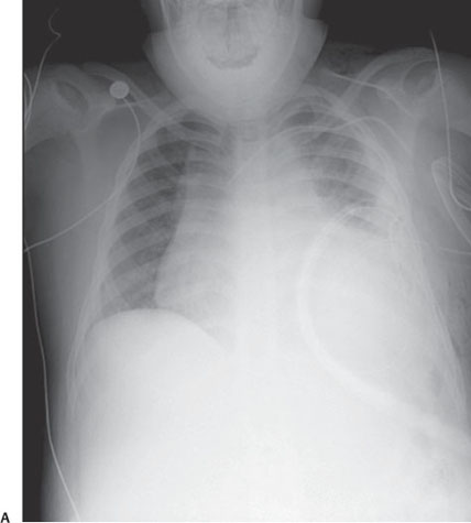
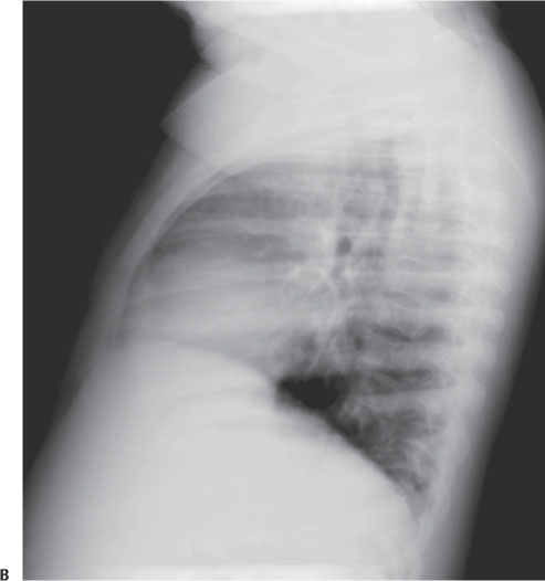
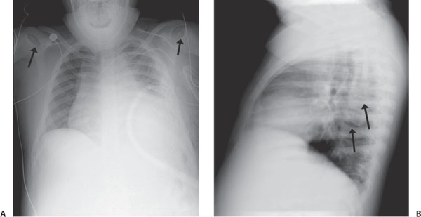
Case 4 A 10-year-old boy with chest pain. A follow-up study was obtained after the acute episode. (A) Frontal chest radiograph demonstrates cardiomegaly and left lower lobe air space disease. There is irregularity of the humeral head epiphyses (arrows). (B) Lateral chest radiograph: there are multiple vertebral body end plate irregularities (arrows). • Acute chest syndrome (ACS), sickle cell disease:
Clinical Presentation
Further Work-up
Imaging Findings
Differential Diagnosis
![]()
Stay updated, free articles. Join our Telegram channel

Full access? Get Clinical Tree


