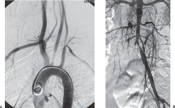
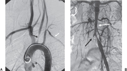
Case 4 The patient is a 13-year-old girl with a diagnosis of “vascular disease.” (A) Digital subtraction aortogram in the left anterior oblique view demonstrates a smooth decrease in caliber of the left common carotid artery (black arrow) and diffuse smooth stenosis of the left subclavian artery (white arrow). (B) The distal aorta shows smooth tapering below the renal arteries (white arrow). There is marked hypertrophy of the lumbar arteries. The right common iliac artery is occluded (black arrow).

 Clinical Presentation
 Imgaging Findings

 Differential Diagnosis
Stay updated, free articles. Join our Telegram channel

Full access? Get Clinical Tree


