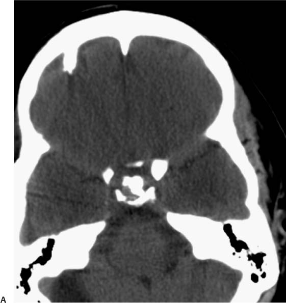
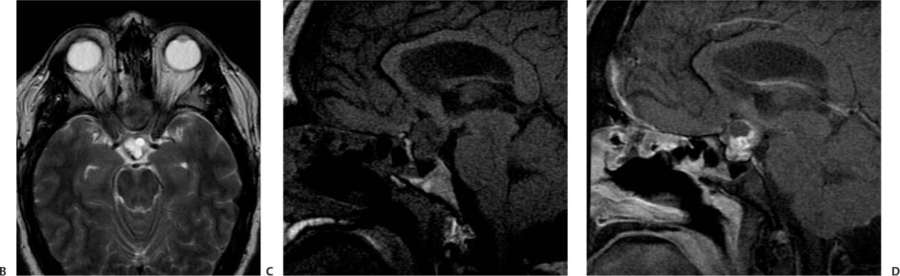
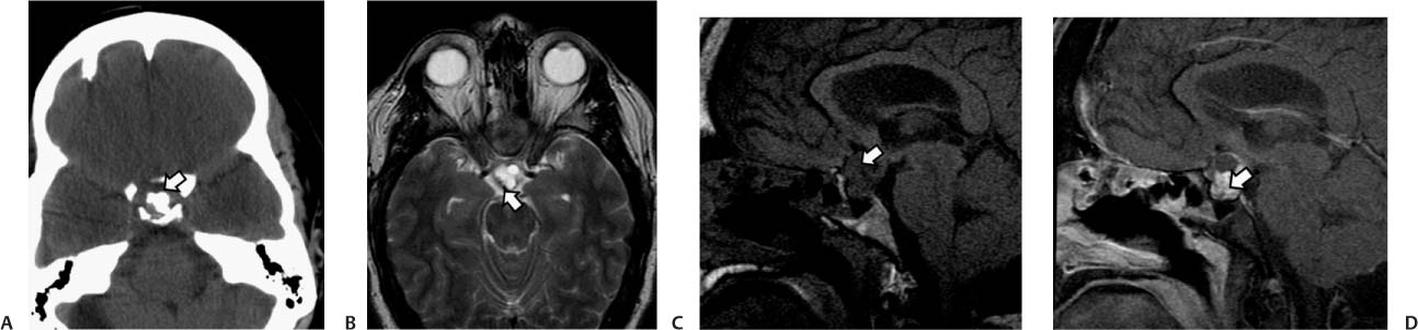
Case 40 A 40-year-old woman with severe headache and blurry vision. (A) Axial computed tomography scan of the brain shows irregular calcifications in the supraclinoid region (arrow). (B) On axial T2-weighted image (WI), the lesion has a multiloculated appearance. Note the dark posterior rim arising from calcification (arrow). (C) Sagittal T1WI shows sellar and suprasellar components of the mass, which is heterogeneous in signal (arrow). (D) Sagittal T1WI after gadolinium injection shows enhancement in the solid areas of the tumor (arrow) and nonenhancing cystic areas. • Craniopharyngioma:
Clinical Presentation
Further Work-up
Imaging Findings
Differential Diagnosis
![]()
Stay updated, free articles. Join our Telegram channel

Full access? Get Clinical Tree


