 Clinical Presentation
A 43-year-old man with chest pain after a motor vehicle accident.
 Imaging Findings
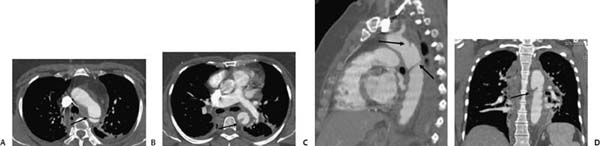
(A–D) Contrast-enhanced thoracic computed tomography (CT): Axial images (A, B) and sagittal and coronal re-formations (C, D) demonstrate an abnormal appearance of the distal aortic arch and proximal descending aorta, an intimal flap, and abnormal change in the aortic caliber (arrows). Abnormal density of the mediastinum, consistent with mediastinal hematoma, is noted.
 Differential Diagnosis
• Traumatic aortic injury (TAI): In approximately 90% of cases, TAI occurs in the region of the aortic isthmus, just distal to the origin of the left subclavian artery. The combination of a mediastinal hematoma in contact with the aorta and an irregularity of the aortic wall resulting from a pseudoaneurysm or an intimal flap confirms the diagnosis.
Stay updated, free articles. Join our Telegram channel

Full access? Get Clinical Tree


