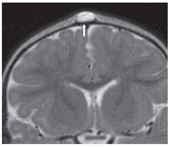
FINDINGS Figure 40-1. Sagittal T1WI. There is a well-circumscribed T1 hypointense subgaleal mass over the anterior fontanel (arrow). Figure 40-2. Coronal T2WI through the mass. The mass is hyperintense (arrow). The lesion did not restrict diffusion nor contrast enhance (not shown).
DIFFERENTIAL DIAGNOSIS Sebaceous cyst, epidermoid cyst, dermoid cyst.
DIAGNOSIS Subgaleal dermoid cyst.
DISCUSSION
Stay updated, free articles. Join our Telegram channel

Full access? Get Clinical Tree


