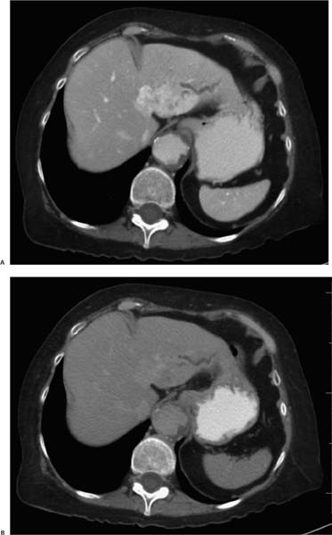
Case 40 A 72-year-old woman with a known history of abdominal aortic aneurysm presents with nausea, vomiting, and jaundice. (A) Contrast-enhanced computed tomography (CT) shows avid arterial-phase enhancement of a single large mass in the left lobe of the liver, indistinctly marginated (large arrow) and causing intrahepatic bile duct dilatation (small arrow). Known aortic aneurysm is noted. (B) Delayed imaging (15 minutes) shows persistent mild enhancement (arrow) of the mass compared with the adjacent liver parenchyma. • Intrahepatic cholangiocarcinoma (ICC): This is strongly indicated by a single large, indistinctly marginated mass showing persistent enhancement on delayed imaging; intrahepatic duct dilatation supports this diagnosis.

 Clinical Presentation
 Imaging Findings

 Differential Diagnosis
Stay updated, free articles. Join our Telegram channel

Full access? Get Clinical Tree


