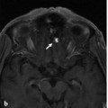
Fig. 40.1 Sagittal T2-weighted (a,b) and T1-weighted (c,d) MRIs demonstrate hematomyelia with a localized blood clot in the center of the cord at the C2 level and no dilated perimedullary vessels. Blood was seen to extend both cranially and caudally along the central canal. The patient was first treated conservatively. After he regained consciousness, conventional angiography was performed to rule out the remote possibility of a microarteriovenous malformation. Case continued in ▶ Fig. 40.2.
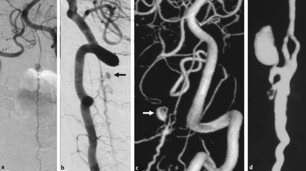
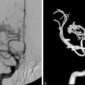
Fig. 40.2 Left vertebral artery angiograms in anteroposterior (AP) (a) and lateral (b) views and 3D rotational reconstruction (c,d) visualized a spinal perimedullary arteriovenous fistula fed by a slightly enlarged anterior spinal artery that arose from the left vertebral artery. At the C2 level, there was a focal unfused segment of the anterior spinal artery (ASA). The fistula was selectively supplied by one of the two unfused limbs of the ASA at the level of C2 with a venous (false) aneurysm (arrows) as the point of rupture. Case continued in ▶ Fig. 40.3.
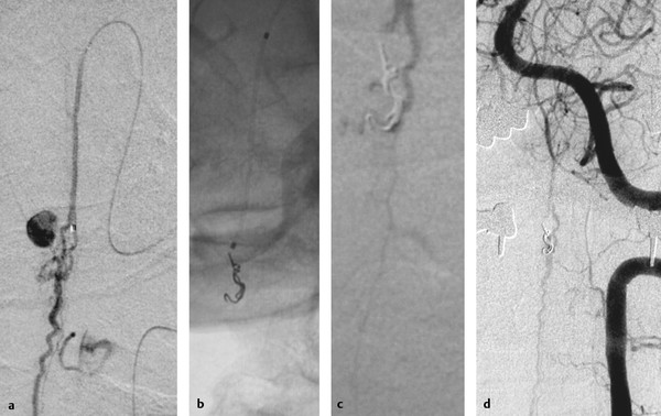
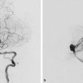
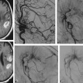
Fig. 40.3 Selective microcatheter injection before (a), plain radiography during (b), and vertebral artery angiograms after (c,d) the embolization demonstrate deposition of two microcoils that led to occlusion of the shunt, while the ASA was preserved.
40.1.3 Diagnosis
Spinal perimedullary arteriovenous fistula arising from an unfused segment of the anterior spinal artery.
40.2 Anatomy and Embryology
Although in the embryo each radicular artery gives rise to a radiculomedullary artery to supply the spinal cord, in postnatal life, the number of radicular arteries supplying the spinal cord is reduced after a transformation and fusion process. At a few unpredictable segmental levels, the radicular artery has a persistent supply to the cord that reaches either the anterior surface via the ventral nerve root or the posterolateral surface via the dorsal nerve root to form and supply the superficial spinal cord arteries. Two to 14 (on average, six) anterior radiculomedullary arteries persist as the result of this “pruning process” of the feeding vessels. The posterior radiculomedullary arteries are reduced less drastically, mostly to between 11 and 16 vessels.
Several nomenclatures and classifications have been used to describe spinal cord arteries, which may lead to some confusion. The original classification is based on where the spinal cord arteries run (i.e., on the posterior, posterolateral, or anterior surface of the spinal cord, thereby constituting the posterior or posterolateral arteries and the anterior spinal artery). A classification proposed by Lasjaunias differentiates three types of spinal radicular arteries according to their region of supply: radicular, radiculopial, and radiculomedullary.
The spinal radicular artery is a small branch, present at every segmental level, that supplies the nerve root as well as the adjacent dura. The radiculopial arteries supply the nerve root and the dorsolateral superficial pial system (via the posterior radicular artery). The radiculomedullary arteries supply the nerve root, the superficial pial system, and the medulla (via the anterior radicular artery). This classification offers advantages for the interventional neuroradiologist when compared with the classical differentiation because it stresses the importance of the anterior supply for the gray matter of the spinal cord parenchyma. Because the anterior spinal artery has radicular, radiculopial, and medullary supply and the radiculopial posterolateral arteries may have some medullary supply (i.e., part of the posterior horns), this classification may, however, result in misunderstandings. This is why we proposed recently the following slight modification to the classification to overcome the anatomical confusions: Radicular arteries are vessels that supply the nerve root and the dura mater but do not supply the spinal cord. These arteries are present on every single segmental level, whereas the following two types may or may not be present at a given segmental level.
First is the anterior radiculomedullary artery, which runs with the anterior nerve root to join the longitudinal trunk on the anterior surface of the spinal cord (i.e., the anterior spinal artery). In addition to this main supply, a minor lateral contribution to a superficial pial network is also derived from the anterior radiculomedullary artery.
Second is the posterior radiculomedullary artery, which accompanies the posterior nerve root and joins the longitudinal systems of posterolateral and/or posterior spinal arteries. The first one is lying laterally, the second one medial to the posterior nerve root entry zone. These arteries mainly supply the pial superficial network of vessels but may give small branches to the gray matter of the posterior horns. One has to bear in mind that these latter arteries predominantly supply the surface of the spinal cord (i.e., the white matter), whereas the anterior radiculomedullary arteries predominantly supply the gray matter of the spinal cord (▶ Fig. 40.4).
Stay updated, free articles. Join our Telegram channel

Full access? Get Clinical Tree


