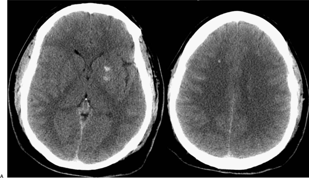
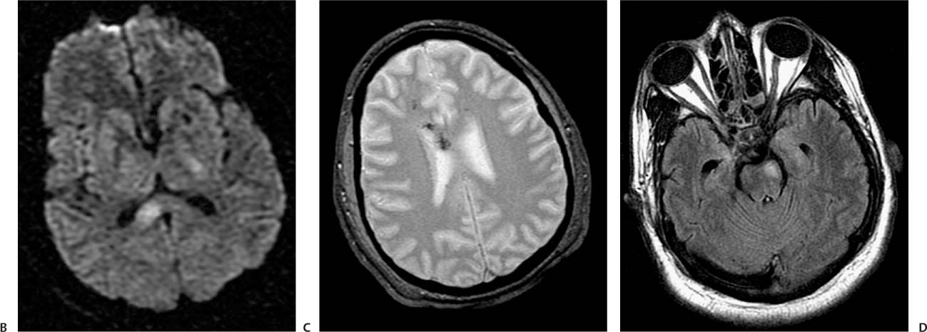
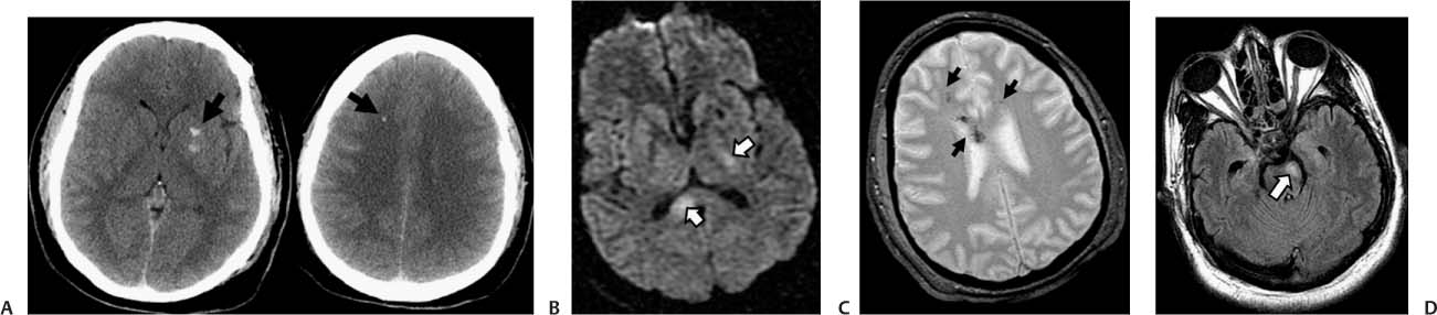
Case 41 A young woman presenting with loss of consciousness after being involved in a motor vehicle collision. (A) Computed tomography (CT) scan of the brain demonstrates multiple punctate foci of hemorrhage in the left basal ganglia and right frontal deep white matter (arrows). (B) Diffusion-weighted image (WI) shows restricted diffusion in the left basal ganglia and in the splenium of the corpus callosum (arrows). (C) Petechial hemorrhage in the deep white matter and corpus callosum is evident in the gradient-echo (GRE) T2*WI (arrows). Multiple other foci were demonstrated in the brainstem and centrum semiovale. (D) Fluid-attenuated inversion recovery image shows increased signal in the left cerebral peduncle (arrow). • Diffuse axonal injury (DAI):
Clinical Presentation
Further Work-up
Imaging Findings
Differential Diagnosis
![]()
Stay updated, free articles. Join our Telegram channel

Full access? Get Clinical Tree


