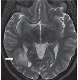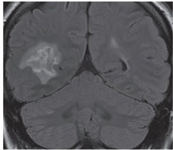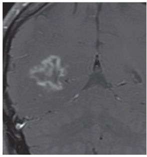


FINDINGS Figure 41-1. Right parasagittal T1WI MRI of the brain showing a heterogeneous hypointense lesion in the right occipito-temporo-parietal white matter (WM) with very minimal mass effect (arrow). Figure 41-2. Axial T2WI through the mass. The mass (arrow) is lateral to the right trigone and occipital horn showing shades of hyperintensity from central almost cerebrospinal fluid (CSF) intensity to less hyperintense variegated heterogeneous periphery. There is no significant mass effect. Figure 41-3. Coronal FLAIR through the lesion showing a three-layer pattern of isointense irregular core with surrounding irregular crinkled hyperintensity and a periphery of smudgy medium hyperintensity presumably edema. Figure 41-4. Coronal post-contrast T1WI. There is a 3.3 cm × 2 cm irregular thick crinkled avid enhancement corresponding to the middle layer in Figure 41-3 surrounding an irregular iso/hypointense core. The peripheral portion is isointense with the surrounding brain. The axial post-contrast images (not shown) show some discontinuity in the enhancing portion. There were a few non-contrast-enhancing periventricular focal T2 hyperintensity elsewhere in the bilateral cerebral WM (not shown).
Stay updated, free articles. Join our Telegram channel

Full access? Get Clinical Tree


