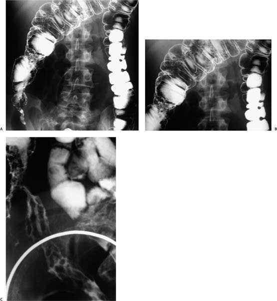
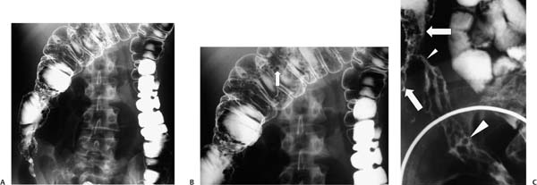
Case 41 A 23-year-old man presents to the gastroenterology clinic with abdominal pain. (A) Double-contrast barium enema shows a cone-shaped cecum and multiple polypoid lesions in the ascending and transverse colon. (B) Magnified view shows small, well-circumscribed polypoid lesions (arrow). (C) Compression view of the terminal ileum and cecum shows nodular, thickened ileal folds (large arrowhead); stricturing of the distal terminal ileum (small arrowhead); and a cone-shaped, nodular cecum (arrows). • Crohn disease (CD): The constellation of multiple inflammatory filiform polyps; a cone-shaped, strictured cecum; and a strictured, nodular terminal ileum is most likely caused by CD. • Lymphoma: This may be indistinguishable from CD in the terminal ileum and cecum but is less likely to cause luminal narrowing. • Tuberculosis:
 Clinical Presentation
 Imaging Findings
 Differential Diagnosis
![]()
Stay updated, free articles. Join our Telegram channel

Full access? Get Clinical Tree


