CASE 41
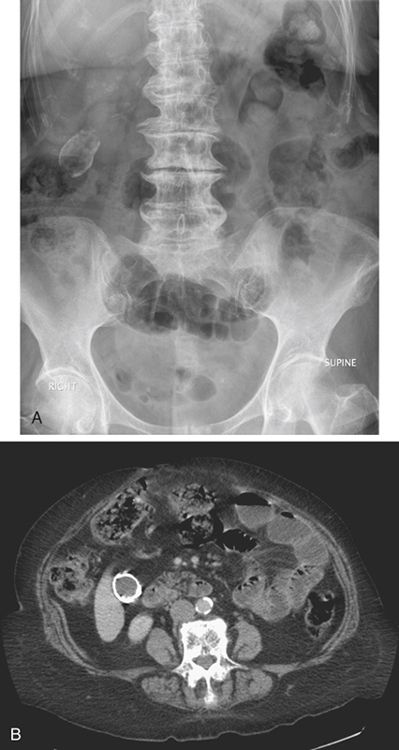
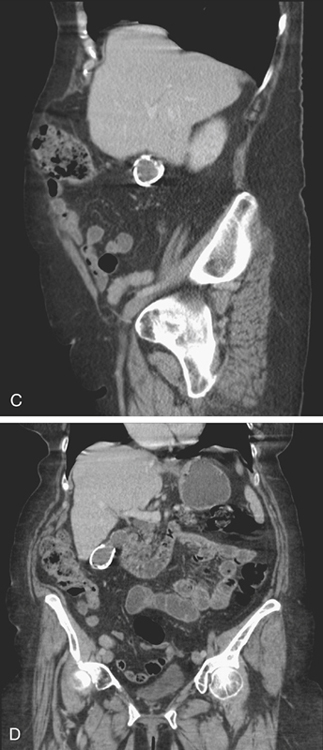
History: An 80-year-old woman presents with left-sided abdominal pain and vomiting.
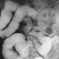
1. What is your differential for the location and diagnosis of the large calcification seen in the right upper quadrant of the abdomen in figure A? (Choose all that apply.)
A. Adrenal gland; e.g., tuberculosis
B. Gallbladder; e.g., porcelain gallbladder
D. Kidney; e.g., calcified cyst
E. Abdominal wall; e.g., old hematoma
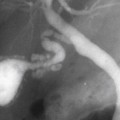
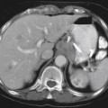
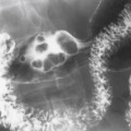
2. Based on the figures, which of the following is the most likely diagnosis?
Stay updated, free articles. Join our Telegram channel

Full access? Get Clinical Tree


