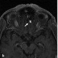
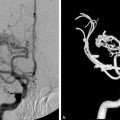
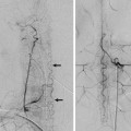
Fig. 41.1 Sagittal T2-weighted (a) and T1-weighted (b) MRIs demonstrate hematomyelia, cord edema, and posteriorly located perimedullary flow voids that enhance after contrast administration in the mid and lower thoracic region, raising suspicion of a spinal cord AVM. Left T10 segmental artery angiograms in AP (c) and lateral (d) views reveal a shunting lesion that was fed by both the anterior (arrow) and the posterior (white arrow) spinal arteries. As an anatomical variation, the supply to both the anterior and the posterior axis arose from the same segmental artery. An aneurysm that is located in the midline and centrally within the cord is noted as the rupture point. Case continued in ▶ Fig. 41.2.
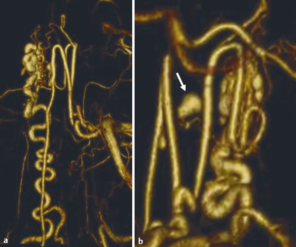
Fig. 41.2 3D rotational reconstruction images (a,b) demonstrate that the aneurysm (arrow) arose from a sulcocommissural artery that interconnects the anterior and the posterior spinal arteries.
41.1.3 Diagnosis
Pial nidus-type arteriovenous malformation (AVM) with a false aneurysm arising from a sulcocommissural artery of the anterior spinal artery.
41.2 Anatomy
The main source of arterial supply to the cord is the anterior spinal artery (ASA) on its ventral axis. The arteries directly supplying the spinal cord that arise from the ASA are the central (sulcal and sulcocommissural) arteries that originate from the anterior spinal artery and the perforating branches arising from the superficial pial network, covering the spinal cord. Sulcal arteries are centrifugal, have a diameter between 0.1 and 0.25 mm, and supply the largest part of the gray matter. They penetrate the parenchyma to the depth of the anterior fissure, course to one side of the cord, and branch mainly within the gray matter (▶ Fig. 41.3).
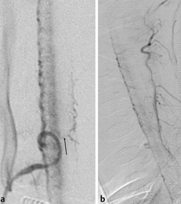
Fig. 41.3 Spinal angiograms in lateral view in two patients (a,b) with cord hyperemia demonstrate the sulcocommissural arteries that supply the anterior aspect of the cord. The arrow points to a transmedullary anastomosis that connects the anterior axis to the posterior axis through the cord.
Stay updated, free articles. Join our Telegram channel

Full access? Get Clinical Tree


