 Clinical Presentation
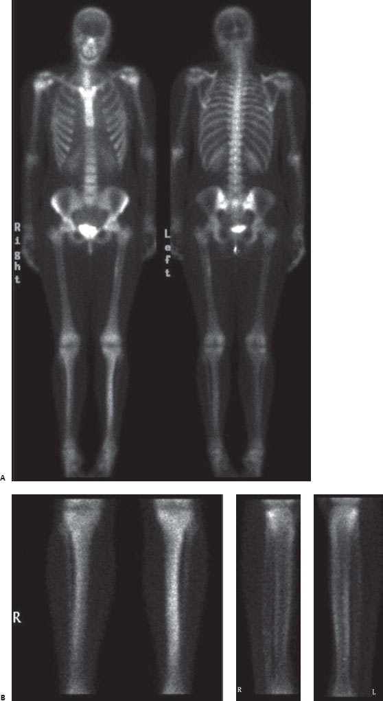
A 19-year-old woman with recent left leg pain.

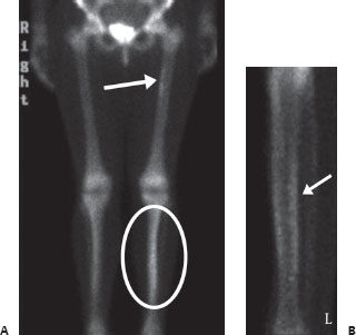
(A) Anterior and posterior whole-body bone scan images demonstrate linear increased uptake along the medial cortical surface of the left proximal femur (arrow). Additionally, there is less-well-defined increased uptake at the mid tibiae, the left greater than the right (circle). (B) Anterior and lateral spot views of the tibiae/fibulae demonstrate that the increased uptake is most pronounced along the posterior cortical surfaces of the tibiae, the left greater than the right, in a linear distribution (arrow).
 Differential Diagnosis
• Shin and hip “splints”: Linear uptake along the posterior periosteal surface of the mid tibia is essentially pathognomonic for shin splints. A similar appearance can be seen along the medial surface of the proximal femur.
• Hypertrophic osteoarthropathy:


Stay updated, free articles. Join our Telegram channel

Full access? Get Clinical Tree


