 Clinical Presentation
A 35-year-old morbidly obese woman with chest pain after a motor vehicle accident.
 Imaging Findings
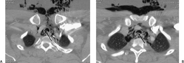
(A, B) Contrast-enhanced computed tomography (CT) of the chest shows significant subcutaneous and mediastinal emphysema, as well as irregularity and a thin, linear, air-density band in relation to the posterior tracheal wall (arrows).
 Differential Diagnosis
• Tracheal rupture: Deformity and discontinuity of the tracheal wall associated with pneumomediastinum are consistent with tracheal rupture.
• Pneumomediastinum without tracheal rupture:
Stay updated, free articles. Join our Telegram channel

Full access? Get Clinical Tree


