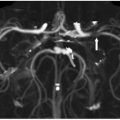
FINDINGS Figure 42-1. F-18 FDG-PET. Top two rows: brain surface project hypermetabolic maps. Bottom two rows: hypometabolic maps. There is markedly decreased metabolism (yellow areas) in bilateral frontal lobes and moderately decreased metabolism (blue areas) in occipital, temporal, and parietal lobes. The occipital lobe hypometabolism is characteristic of Lewy body disease.
DIFFERENTIAL DIAGNOSIS Alzheimer disease (AD), Dementia with Lewy bodies (DLBs). Parkinson disease with dementia (PDD), normal pressure hydrocephalus (NPH), Parkinson-plus syndromes (progressive supranuclear palsy, multisystem atrophy, corticobasal degeneration).
DIAGNOSIS Dementia with Lewy bodies (DLBs).
DISCUSSION
Stay updated, free articles. Join our Telegram channel

Full access? Get Clinical Tree


