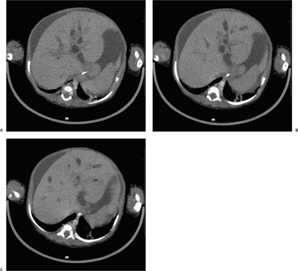
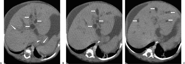
Case 42 A 12-month-old boy presents with jaundice. (A–C) Infused abdominal computed tomography (CT) shows multiple cystic structures (arrows) throughout the liver parenchyma, from the hilum to the periphery, in the general distribution of the bile ducts. Some of these structures may contain stone or sludge. The common bile duct is also dilated. Ascites is noted (arrowheads). • Caroli disease: This is the most likely diagnosis for saccular and cystic structures within the liver parenchyma extending to the porta hepatis. • Biliary obstruction: This typically causes uniform peripheral biliary dilatation rather than this focal, saccular appearance (from congenital causes such as biliary atresia). • Polycystic liver disease:
 Clinical Presentation
 Imaging Findings
 Differential Diagnosis
![]()
Stay updated, free articles. Join our Telegram channel

Full access? Get Clinical Tree


