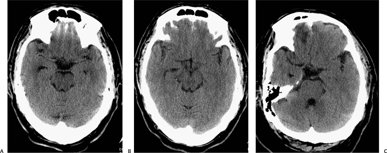
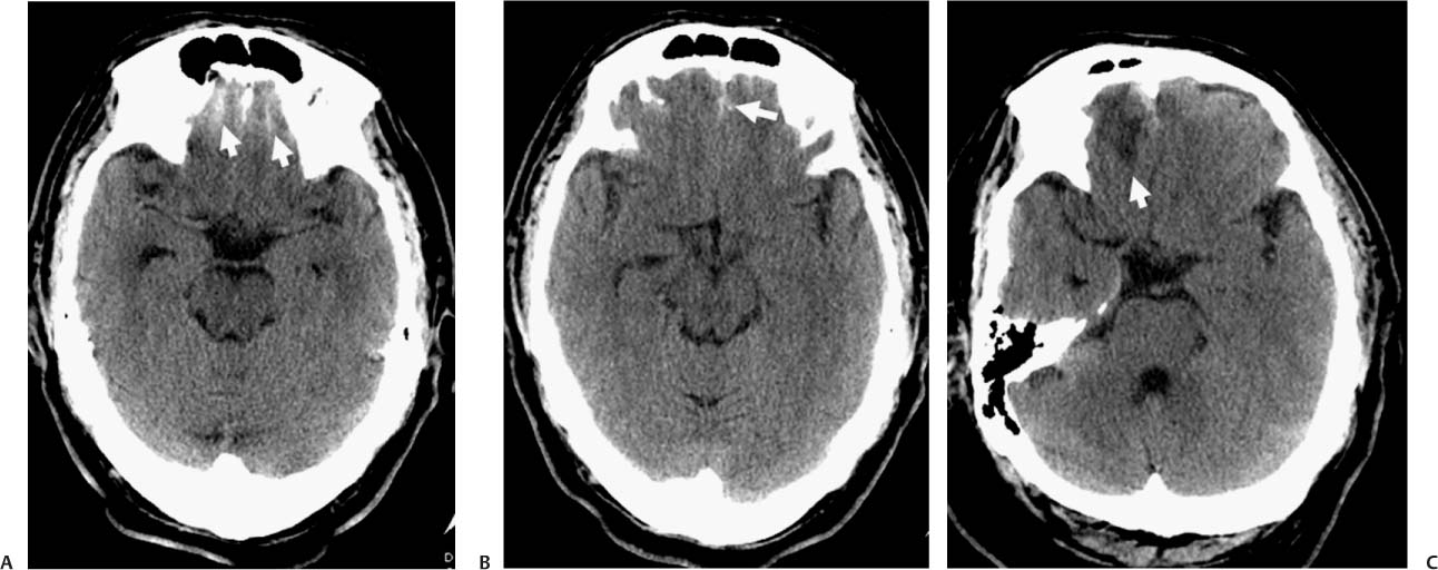
Case 43 A 35-year-old with headaches following an assault. (A) Computed tomography (CT) demonstrates increased attenuation along the inferior frontal sulci (arrows). No fracture was identified in the images with bone algorithm. (B) CT demonstrates increased attenuation in the interhemispheric region (arrow). (C) Follow-up CT 2 days later shows low attenuation in the right frontal lobe inferiorly, consistent with edema (white arrow). The hemorrhage is still visible. • Traumatic subarachnoid hemorrhage (SAH): On CT, this shows high-attenuation blood within the basal cisterns and subarachnoid spaces. Traumatic hemorrhage is located in the convexities more frequently than in the basal cisterns. Nonvisualization of the interpeduncular cistern may be a clue that a small amount of isodense subarachnoid blood is present.
Clinical Presentation
Imaging Findings
Differential Diagnosis
Stay updated, free articles. Join our Telegram channel

Full access? Get Clinical Tree


