 Clinical Presentation
A 53-year-old woman with a past medical history of anticoagulation therapy for pulmonary embolism and deep vein thrombosis, now presenting with acute chest pain.
 Imaging Findings
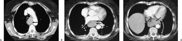
(A–C) Contrast-enhanced computed tomography (CT) of the chest: axial images demonstrate a large, well-defined hyperdensity throughout the entire length of the esophageal wall, eccentrically located and displacing the lumen anteriorly and to the right (arrows).
 Differential Diagnosis
• Intramural hematoma of the esophagus (IHE): Acute IHE may present as a hyperdense collection located either concentrically or eccentrically in the esophageal wall.
• Esophageal tumor:
Stay updated, free articles. Join our Telegram channel

Full access? Get Clinical Tree


