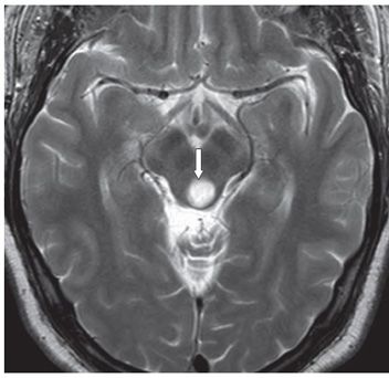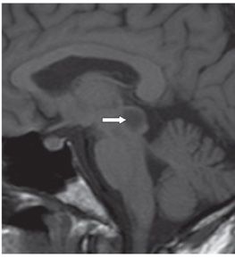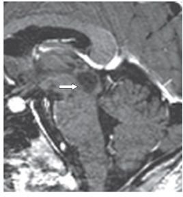


FINDINGS Figures 43-1 and 43-2. Axial FLAIR and corresponding T2WI respectively through the midbrain. There is a 1.2-cm round hyperintense dorsal midbrain mass (arrows) encompassing the cerebral aqueduct. Apart from a mild bulge posteriorly, there is no mass effect. Figures 43-3 and 43-4. Sagittal pre- and post-contrast T1WI through the mass. The mass is hypointense and does not enhance (arrows). There is a posterior bulge of the colliculi with apparent obliteration of the aqueduct.
DIFFERENTIAL DIAGNOSIS Tectal glioma, pineal mass, midbrain cyst, neurofibromatosis type 1 (NF1).
DIAGNOSIS
Stay updated, free articles. Join our Telegram channel

Full access? Get Clinical Tree


