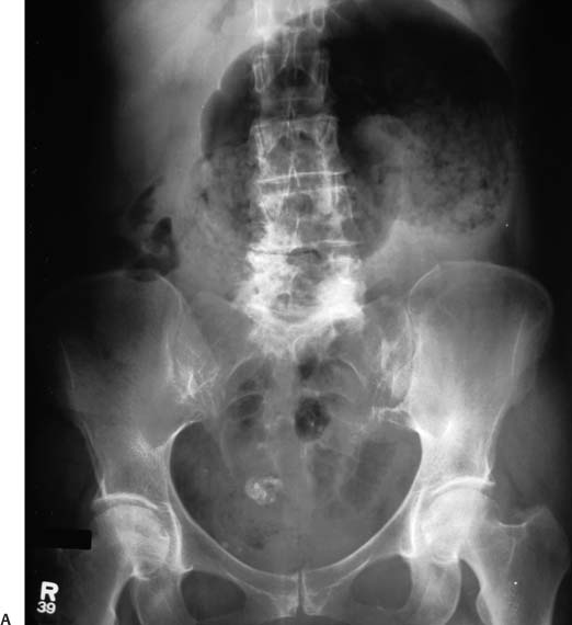
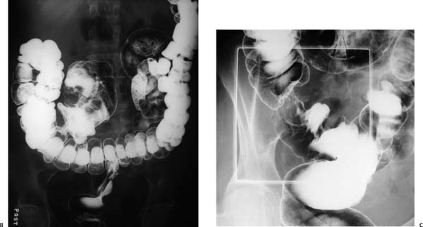
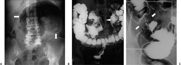
Case 43 A 55-year-old woman presents with intense lower abdominal pain and distension. (A) Supine frontal abdominal radiograph shows markedly dilated middle to left bowel loop (arrows) with a caudal cleft, giving it the appearance of a coffee bean. No other dilated bowel is noted. (B) Single-contrast barium enema shows a dilated cecum projecting over the midline (arrowheads). After the radiograph was obtained, cecal dilatation diminished. (C) Compression view shows an apple-core lesion, consistent with colon carcinoma (arrows). • Cecal volvulus caused by colon carcinoma: This is the only possible diagnosis, given the cecal obstruction, the resolution before or during the barium enema, and the apple-core stricture. • Sigmoid volvulus:
 Clinical Presentation
Further Work-up
 Imaging Findings
 Differential Diagnosis
![]()
Stay updated, free articles. Join our Telegram channel

Full access? Get Clinical Tree


