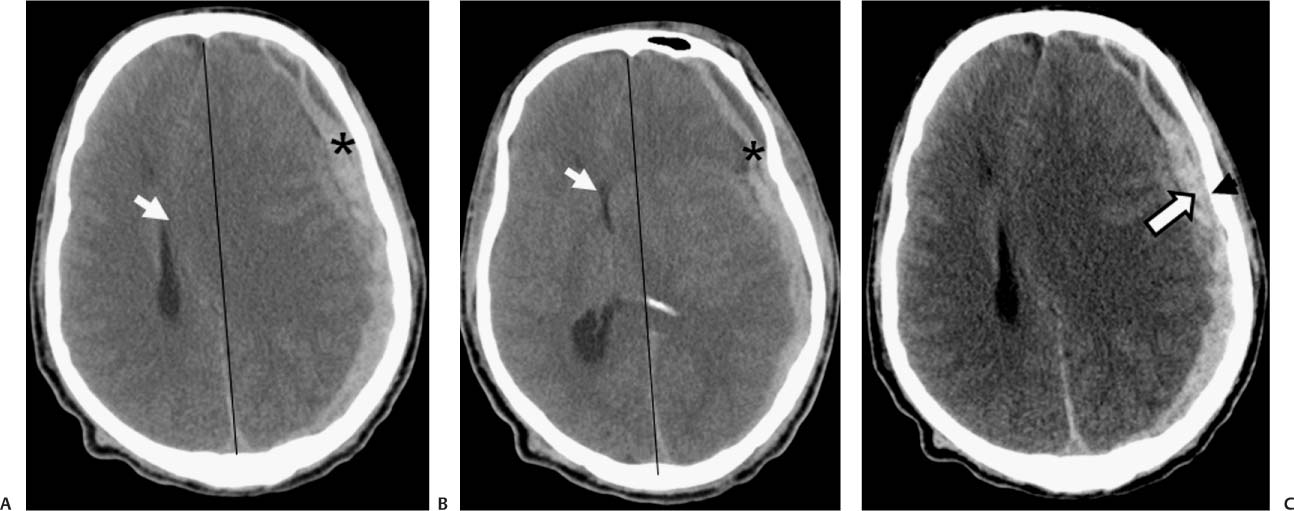
Case 44 A 51-year-old man after a motor vehicle collision. (A,B) Computed tomography (CT) scans with “brain windows” reveal a crescent-shaped extra-axial collection with high attenuation that is slightly heterogeneous (asterisk). It compresses the left lateral ventricle and causes rightward deviation of the midline structures (arrow). (C) Note the increased contrast between the hematoma (arrow) and the skull (arrowhead) in the “blood” window. • Acute subdural hematoma (SDH): Acute SDH is a crescentic, hyperdense collection between the dura and arachnoid membrane. Displacement of the cortex away from the inner table is common. It can cause midline shift with compression of the ipsilateral lateral ventricle and dilatation of the contralateral ventricle. • Epidural hematoma:
Clinical Presentation
Imaging Findings
Differential Diagnosis
![]()
Stay updated, free articles. Join our Telegram channel

Full access? Get Clinical Tree


