 Clinical Presentation
A 25-year-old pregnant woman with acute shortness of breath. Chest radiograph was normal.

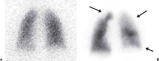
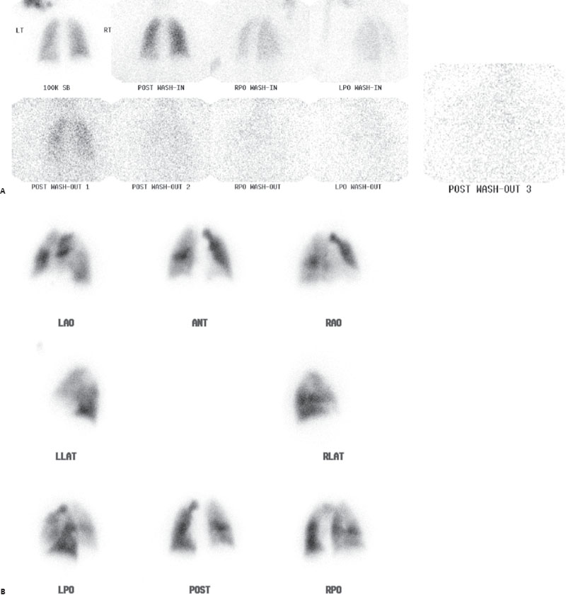
(A) Sequential posterior and posterior oblique xenon 133 ventilation images are normal. There are no defects on single-breath or early wash-in imaging, and there is no abnormal retention on washout. (B) Tc99m-MAA images demonstrate multiple medium and large subsegmental and segmental perfusion defects (arrows)—some are decreased and others are completely absent.
 Differential Diagnosis
• High probability of pulmonary embolism (PE): At least two large (or the equivalent) mismatched perfusion defects meet the criteria for high probability.
• Pulmonary vasculitis: This is also in the differential for multiple mismatches but is rare and less likely given the history.
• Mediastinal adenopathy compressing pulmonary vessels: This is also in the differential for mismatches but usually does not result in this many widespread defects; furthermore, it is unlikely, given the normal chest x-ray (CXR).


Stay updated, free articles. Join our Telegram channel

Full access? Get Clinical Tree


