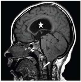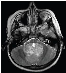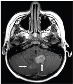


FINDINGS Figure 44-1. Axial NCCT through the posterior fossa. There is a heterogeneous midline posterior fossa mass with scattered calcifications (arrows). Figure 44-2. Sagittal T1WI. The mass is within the fourth ventricle, with extension of tumor through the midline foramen of Magendie (arrow). Notice stretching of the corpus callosum, indicating hydrocephalus (star). Figure 44-3. Axial T2WI through the mass. The lesion is heterogeneous with some cystic foci. Figure 44-4. Axial post-contrast T1WI through the mass. There are areas of enhancement (arrows) within the mass.
DIFFERENTIAL DIAGNOSIS Medulloblastoma, ependymoma, choroid plexus papilloma.
DIAGNOSIS Ependymoma.
DISCUSSION
Stay updated, free articles. Join our Telegram channel

Full access? Get Clinical Tree


