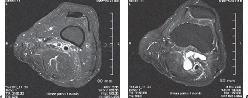
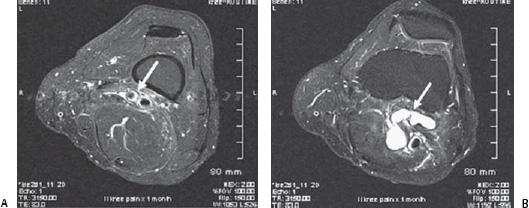
Case 44 The patient is a 50-year-old woman with no previous medical history. (A) Selected enhanced T1-weighted axial image of the right knee demonstrating a rim of high signal intensity surrounding the wall of the popliteal artery (white arrow). The popliteal vein is spared. (B)
 Clinical Presentation
 Imaging Findings
![]()
Stay updated, free articles. Join our Telegram channel

Full access? Get Clinical Tree


