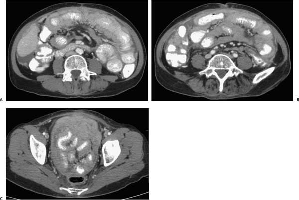
Case 44 A 56-year-old woman presents with nausea, vomiting, and increasing abdominal pain. (A) Contrast-enhanced computed tomography (CT) shows extensive soft tissue infiltrating the mesenteric and omental fat (arrow) in the pelvis and abdomen with marked, diffuse bowel wall thickening. Minimal ascites is noted (arrowhead). (B) More caudal image again shows the infiltrating soft tissue (arrow) as well as peritoneal thickening and nodularity (arrowhead). (C) Pelvic image shows soft tissue (arrows) completely encasing the pelvic structures. • Peritoneal malignant mesothelioma: This is the most likely diagnosis, as indicated by bowel wall thickening with soft-tissue infiltration of the mesenteric and omental fat (caking) and minimal ascites. • Primary peritoneal carcinoma (serous papillary carcinoma):
 Clinical Presentation
 Imaging Findings
 Differential Diagnosis
![]()
Stay updated, free articles. Join our Telegram channel

Full access? Get Clinical Tree


