CASE 44
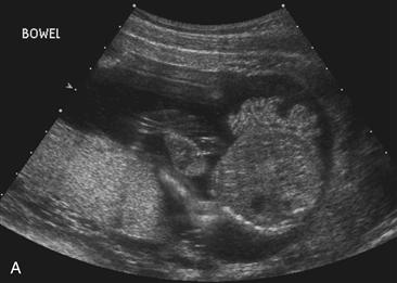
Used with permission from McGahan JP, et al: Fetal abdomen and pelvis. In McGahan JP, Goldberg BB [eds]: Diagnostic Ultrasound, 2nd ed. New York: Informa Healthcare USA, 2008; 1301.
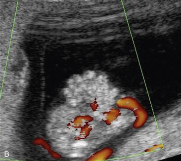
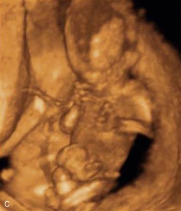
Courtesy of Dolores Pretorius, MD, San Diego, California.
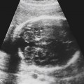
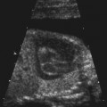
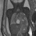
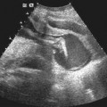
History: A 19-year-old patient presents with an elevated alpha fetoprotein level on screening.
1. What should be included in the differential diagnosis for this 20-week ultrasound scan? (Choose all that apply.)
A. Omphalocele
2. What structure seen on ultrasound is critical in differentiating gastroschisis from omphalocele?
A. Stomach
B. Liver
C. Small bowel
D. Site of umbilical cord insertion
3. Which of the following entities is least likely to be associated with gastroschisis?
D. Small anterior abdominal wall defect
Stay updated, free articles. Join our Telegram channel

Full access? Get Clinical Tree


