Case 45 A 65-year-old with headache and slow, progressive intellectual decline. (A) Computed tomography (CT) at the level of the lateral ventricles shows a crescent-shaped extra-axial collection in the left convexity with high attenuation in the dependent portion (arrow) and fluid attenuation anteriorly (asterisk). (B,C) The slices in the superior aspect of the brain show a large collection (arrows) with layering of blood products causing hematocrit effect. Note the relatively minimal mass effect for the size of the hematoma. • Acute-on-chronic subdural hematoma (SDH): This is characterized by repeated hemorrhage into previous subdural collections. It may show layering of blood products. • Acute SDH:
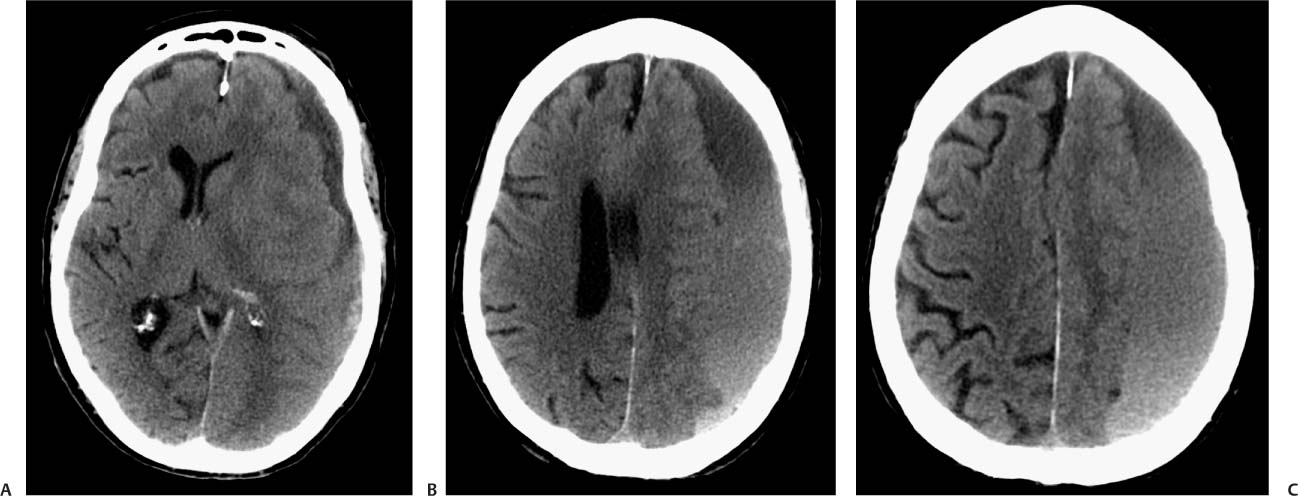
Clinical Presentation
Imaging Findings
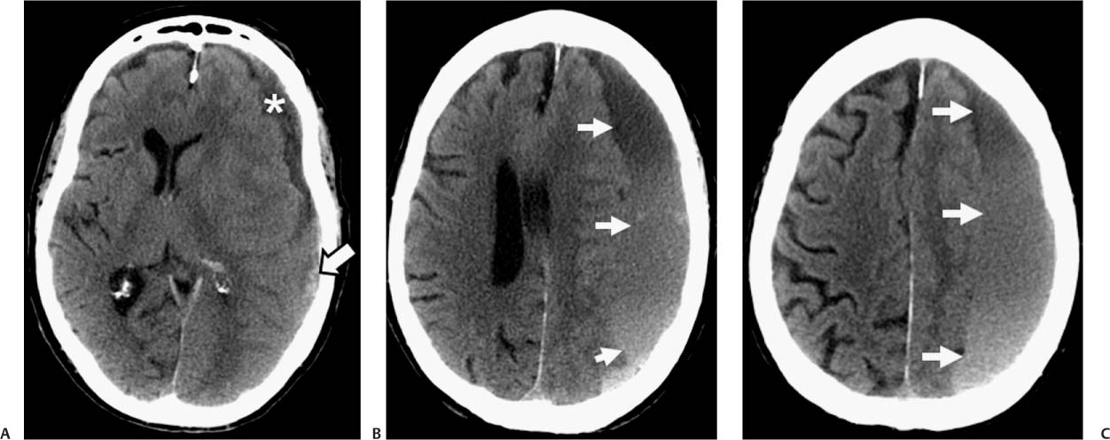
Differential Diagnosis
![]()
Stay updated, free articles. Join our Telegram channel

Full access? Get Clinical Tree


