 Clinical Presentation
A 52-year-old man with dysphagia.
 Imaging Findings
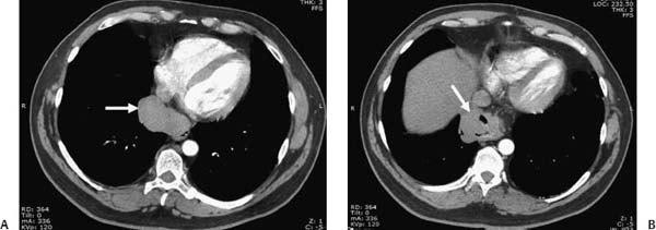
(A, B) Contrast-enhanced thoracic computed tomography (CT) reveals a well-defined soft-tissue-density mediastinal mass eccentrically located in the distal esophagus (arrows). There is a clear interface with adjacent structures with no obvious infiltrative changes.
 Differential Diagnosis
• Esophageal leiomyoma: A well-defined soft-tissue-density mass in the distal esophagus may represent an esophageal leiomyoma.
• Esophageal fibroma/schwannoma: Other benign esophageal tumors (e.g., fibromas and schwannomas) have identical imaging appearances, presenting as noninfiltrative soft-tissue mass.
• Esophageal carcinoma:
Stay updated, free articles. Join our Telegram channel

Full access? Get Clinical Tree


