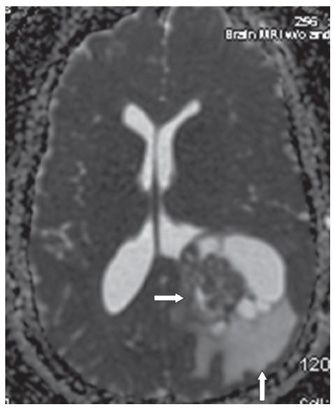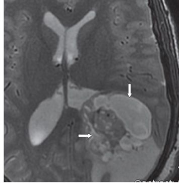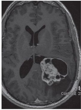


FINDINGS Figures 45-1 and 45-2. Axial DWI with corresponding ADC map through the lateral ventricles. There is a mixed solid cystic mass occupying the left trigone with restricted diffusion in the solid component (transverse arrows). The ADC map shows surrounding parenchymal finger-like hyperintensity consistent with vasogenic edema (vertical arrow in Figure 45-2). Figure 45-3. Axial T2WI. The solid component of the mass is heterogeneous but mostly isointense with gray matter (GM) with tiny hyperintense areas (transverse arrow). The cystic components have cerebrospinal fluid (CSF) intensity (vertical arrow) and posteriorly to the mass is the hyperintense vasogenic edema. Figure 45-4. Axial post-contrast T1WI. There is avid contrast enhancement of the solid component with mild heterogeneity. A rim of contrast enhancement surrounds the entire mass including the cystic components.
DIFFERENTIAL DIAGNOSIS Ependymoma, choroid plexus carcinoma, subependymoma.
DIAGNOSIS Anaplastic ependymoma WHO III.
DISCUSSION
Stay updated, free articles. Join our Telegram channel

Full access? Get Clinical Tree


