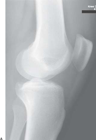
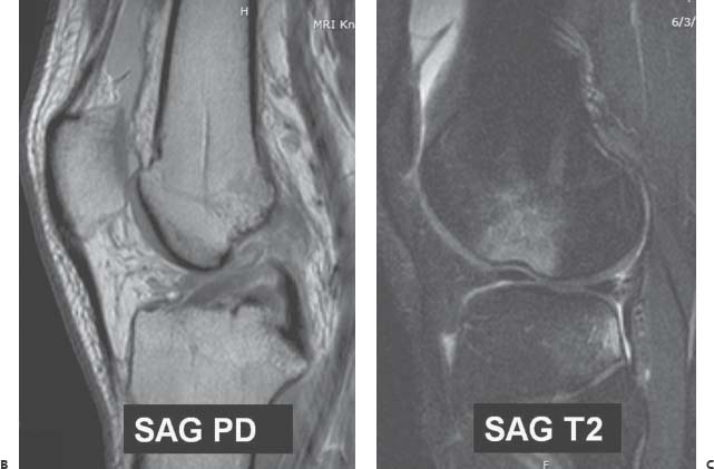
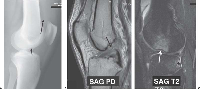
Case 46 The patient is a 19-year-old man who sustained a twisting injury while playing soccer. Further Work-up (A) Radiograph of the knee show an abnormally deep depression over the lateral condylopatellar sulcus (short arrow). A large joint effusion (long arrow) is also present. (B,C) Sagit tal proton density (PD)-weighted and T2-weighted magnetic resonance imaging (MRI) shows complete disruption of the anterior cruciate ligament (ACL: black arrow

 Clinical Presentation

 Imaging Findings

![]()
Stay updated, free articles. Join our Telegram channel

Full access? Get Clinical Tree


