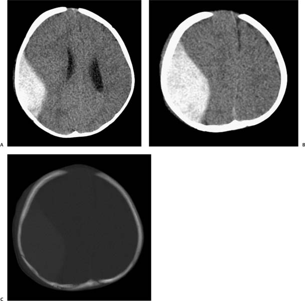
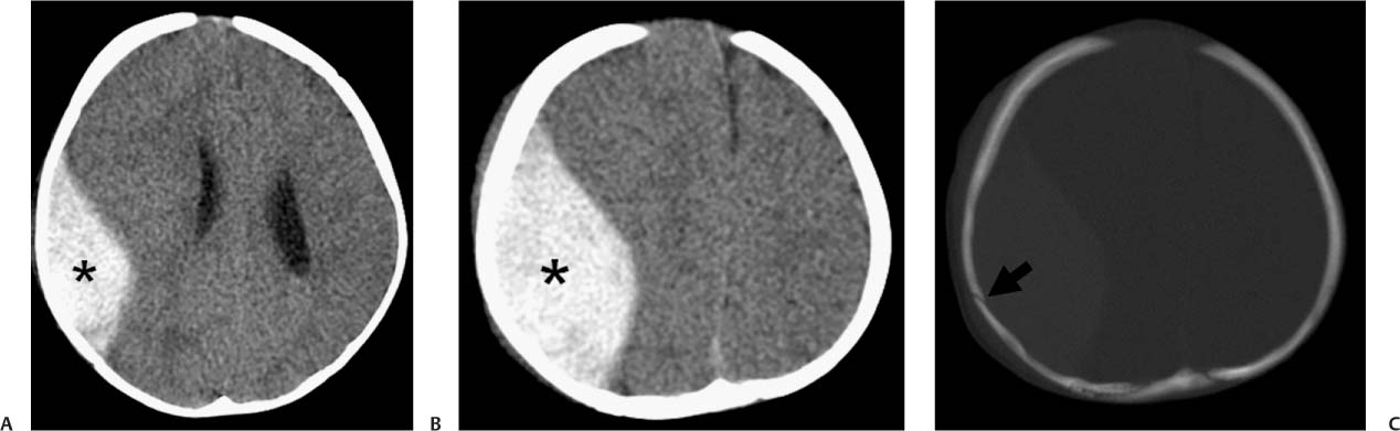
Case 46 A 7-month-old child after a fall. (A,B) Computed tomography (CT) scans of the brain demonstrate a lentiform collection of blood in the right frontoparietal convexity (asterisk). (C) Bone windows demonstrate a linear nondisplaced fracture in the right parietal bone (arrow). • Epidural hematoma (EDH): This is a biconvex, hyperdense extra-axial fluid collection. There is adjacent skull fracture in 90% of cases. On occasion, subdural and epidural hematomas contain alternating crescent-shaped regions of various densities that produce a “swirled” appearance, considered an indication of active hemorrhage. • Subdural hematoma:
Clinical Presentation
Imaging Findings
Differential Diagnosis
![]()
Stay updated, free articles. Join our Telegram channel

Full access? Get Clinical Tree


