46

Case 46


image


image Clinical Presentation


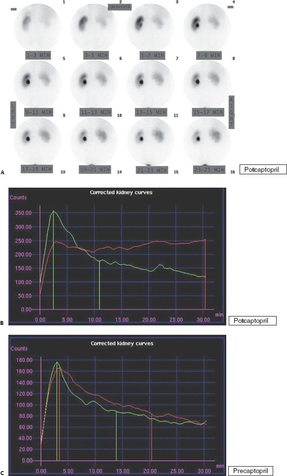
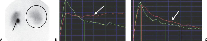
A 42-year-old man with uncontrolled hypertension. Examination was performed post captopril (images and curves shown in Figs. A and B). Baseline examination was normal symmetric (baseline curve shown in Fig. C; right is red, left green; baseline images not shown).


image Imaging Findings


image


(A,B) Posterior images and renogram curves from a Tc99m-MAG3 captopril renal scan demonstrate normal uptake and excretion by the left kidney into an extrarenal pelvis (A, arrow), which progressively clears over time, arguing against obstruction. On the right, there is progressive radiotracer extraction but no demonstrable excretion (A, circle), with resultant flattening of the renogram curve (B, arrow). There is decreased relative right renal function (calculated to be 41% for the right, 59% for the left) as measured at peak uptake at 2.5 minutes (B, asterisks). (C) Renogram curve from the baseline examination before captopril demonstrates normal excretory curves bilaterally (arrow) and symmetric parenchymal function (asterisk).


image Differential Diagnosis


•  Renovascular hypertension (RVH): Unilateral renal parenchymal dysfunction with captopril that normalizes without captopril is highly suggestive of this condition.


•  Chronic medical renal disease:

Stay updated, free articles. Join our Telegram channel

Jan 24, 2016 | Posted by in NUCLEAR MEDICINE | Comments Off on 46

Full access? Get Clinical Tree

Get Clinical Tree app for offline access